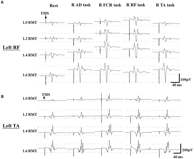
Figure 1. Typical recording of recruitment curves.
Recording of motor evoked potentials from the left rectus femoris (RF) and left tibialis anterior (TA) muscles on a representative subject during different motor tasks of right side limbs. Arrows indicate delivery of transcranial magnetic stimulation (TMS). RMT: resting motor threshold; R: right; AD: anterior deltoid; FCR: flexor carpi radialis; RF: rectus femoris; TA: tibialis anterior.

