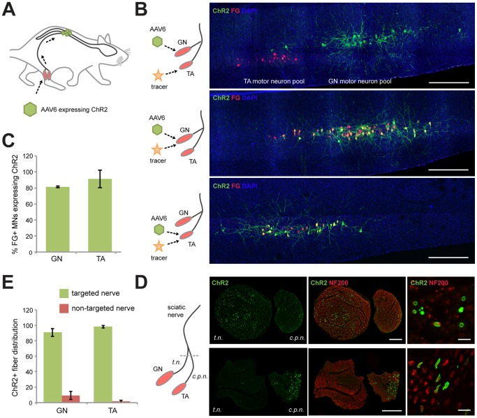
Figure 1. Targeting ChR2 to specific motor neuron axons within the sciatic nerve.
(A) AAV6 encoding ChR2 fused to yellow fluorescent protein (YFP) was injected into the gastrocnemius muscle (GN) or tibialis anterior (TA) muscle, taken up at the neuromuscular junctions and delivered to spinal cord motor neuron (MN) cell bodies via axonal transport. (B) Longitudinal sections of lumbar spinal cord 4 weeks following AAV6 intramuscular injection into GN or TA muscles. The retrograde tracer, Fluoro-Gold (FG), was injected into the ChR2 targeted or non-targeted muscle 1 week prior to analysis. Green, native YFP fluorescence expressed from the ChR2-YFP fusion protein. Red, FG. Blue, DAPI. Scale bar, 1 mm. (C) Percentage of FG positive MNs expressing ChR2 in GN (n = 5) or TA (n = 4). (D) Confocal images of sciatic nerve cross-sections following GN or TA muscle injection. The sciatic nerve bifurcates into the tibial nerve (t.n.) and common peroneal nerve (c.p.n.). Green, ChR2-YFP. Red, Neurofilament 200, a ubiquitous stain for axons. Scale bar, 200 µm. High magnification suggests membrane localization of ChR2. Scale bar, 10 µm. (E) Percentage of total ChR2+ axons in the targeted or non-targeted branches of the sciatic nerve following injection in GN (n = 5) or TA (n = 4).

