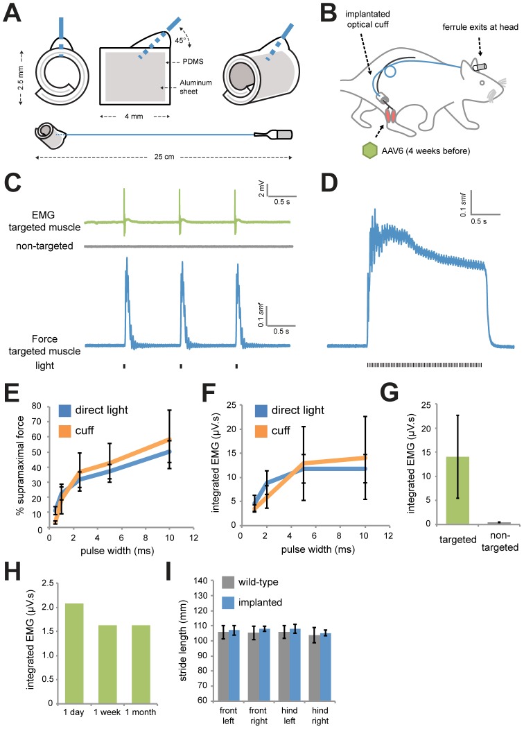
Figure 3. Implantable optical nerve cuffs are well tolerated and activate MNs in anesthetized rats.
(A) Biocompatible spiral cuffs were constructed from polydimethylsiloxane (PDMS) and covalently bound to a silicon-based optical fiber that was terminated with a stainless steel ferrule. (B) Optical nerve cuffs were implanted into rats around the sciatic nerve 4 weeks following AAV6:ChR2 delivery. (C) Typical traces of EMG (targeted and non-targeted muscles) and force (targeted muscle only) following illumination using the optical nerve cuff (20 mW, 5 ms, 1 Hz) in anesthetized rats 1 week following cuff implantation. (D) Representative force trace following a train of light pulses (20 mW, 2.5 ms, 36 Hz) using the nerve cuff. (E) Percentage of smf versus pulse width (20 mW light power) using direct laser light application (n = 7) or light transmitted through the implanted cuff (n = 3). (F) Percentage of smf versus light power (5 ms pulse width) using direct laser light application (n = 7) or light transmitted through the implanted cuff (n = 3). (G) Integrated EMG in targeted and non-targeted muscles following light delivery using the optical nerve cuff (20 mW, 5 ms) (n = 3). (H) Integrated EMG in the targeted muscle at 1 day, 1 week, and 1 month following implantation (n = 1). (I) Stride length of paws in age-matched wild-type littermates (n = 4) and rats 1 week post-implantation of optical nerve cuffs (n = 5).

