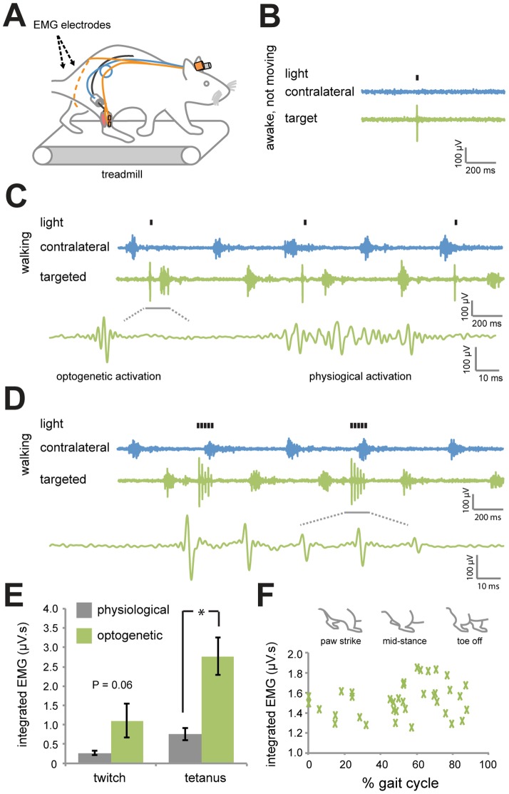
Figure 4. Optogenetic activation of targeted sciatic nerve axons in awake and moving rats.
(A) Optical nerve cuffs and EMG electrodes were implanted into rats 4 weeks following AAV6:ChR2 delivery. EMG electrodes were implanted onto the surface of the AAV6:ChR2-injected muscle (targeted) and onto the uninjected muscle on the opposite leg (contralateral). Non-anesthetized rats were tested for EMG activity on a treadmill 3 days following the surgery. (B) EMG activity in response to a pulse of blue light (20 mW, 5 ms) in the targeted and contralateral muscles in awake, non-moving rats. (C) EMG activity in response to pulses of light (20 mW, 5 ms, 1 Hz) in awake rats walking on a treadmill at constant speed (20 cm/s). (D) EMG activity in response to 150 ms trains of light (20 mW, 5 ms, 36 Hz) in awake rats walking on a treadmill. (E) Integrated EMG in the targeted muscle in response to optogenetic or physiological activation (n = 3, animals matched) for twitch and tetanus contractions. Integrated EMG responses following optogenetic activation are greater or equal to physiological activity (* P<0.05; 2 tailed paired T-test). (F) Integrated EMG versus gait cycle demonstrating that activity was independent of the position of the legs (n = 32 trials, R2 = 0.05).

