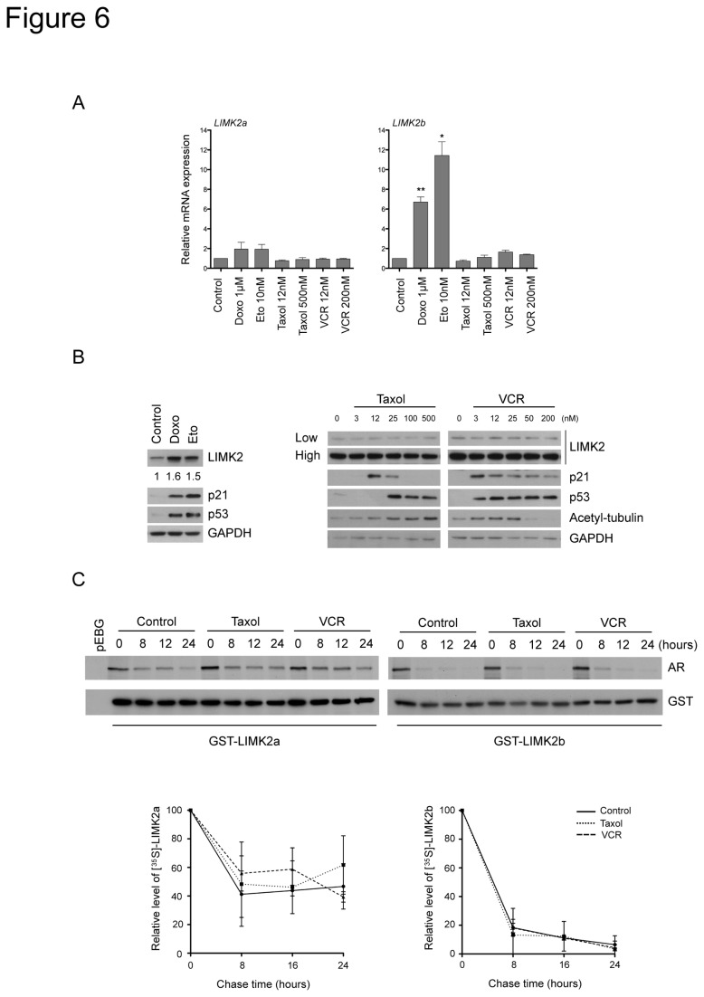
Figure 6. LIMK2 levels are upregulated by DNA damage agents but not by microtubule-targeted drugs.
(A) LIMK2 expression is not regulated at the transcriptional level by microtubule-targeted drugs. SHEP cells were treated with the indicated drugs for 16 hours. The levels of LIMK2a and LIMK2b mRNA were quantified by qRT-PCR, normalized to the housekeeping gene L32, and plotted as relative expression to the control ± S.E.M of three independent experiments (*, p < 0.05; **, p < 0.001, unpaired t-test). (B) LIMK2 protein levels are induced by genotoxic stress but not by treatment with microtubule-targeted drugs. SHEP cells were treated with 1 µM doxorubicin (Doxo), 10 µM etoposide (Eto) or with taxol or vincristine (VCR) at the indicated concentrations for 24 hours before immunoblotting analysis. Two different exposures of LIMK2 immunoblot are shown (low and high). The numbers below the top panel represent the fold-change in the indicated protein levels. (C) Microtubule-targeted drugs do not affect the stability of LIMK2a or LIMK2b proteins. SHEP cells were transiently transfected with GST-LIMK2a or GST-LIMK2b and 24 hours later, they were incubated with 25 nM taxol or 2 nM vincristine (VCR) for 10 hours. Cells were then pulse-labeled with [35S]-methionine/cysteine and chased in the presence of the microtubule-targeted drugs for the indicated time periods. The GST-tagged proteins were purified with glutathione sepharose beads and analyzed by autoradiography (AR) and immunoblotting with anti-GST antibody. Quantification of the level of GST-LIMK2a and GST-LIMK2b proteins from three separate assays is expressed as mean ± S.E.M of percentage values of the samples at zero time.

