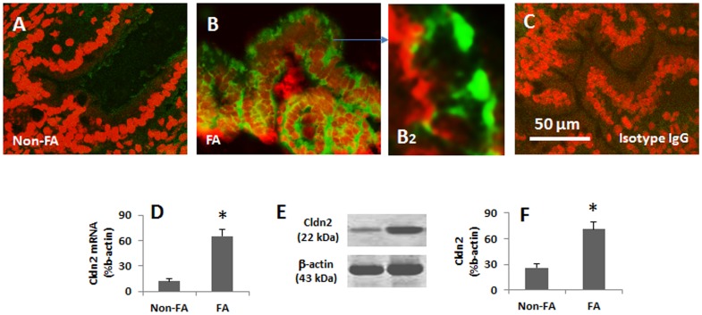
Figure 5. Expression of Cldn2 is increased in gut epithelia of patients with food allergy (FA).
The duodenal biopsies were obtained from 6 patients with food allergy and 6 patients with duodenal peptic ulcers (no FA) and processed for immunohistochemistry. A–C, representative confocal images show the Cldn2 positive staining (in green). C is an isotype control. D, the bars indicate the mRNA levels of Cldn2 in the biopsies that were analyzed by qRT-PCR. E, the immune blots indicate the protein levels of Cldn2 in the biopsies that were analyzed by Western blotting. The β-actin blots were used as an internal control. F, the bars indicate the summarized integrated density of the immune blots in E. The samples from patients were analyzed individually. The data represent 6 separate experiments.

