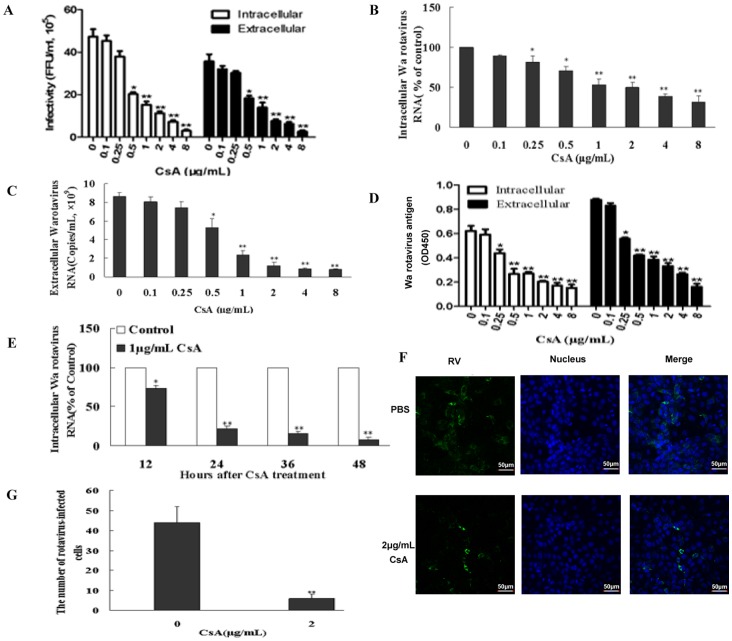
Figure 1. Effect of cyclosporin A (CsA) on Wa rotavirus replication in vitro.
Infectious viral titers (A) were determined by serial dilution and immunofluorescence analysis. The intracellular or extracellular Wa rotavirus is expressed as focus-forming units per milliliter (FFU/mL) of cell lysate or supernatant, respectively. Wa rotavirus RNA expression levels (B) were determined by qRT-PCR. The levels of intracellular Wa rotavirus RNA are expressed as % of control (without CsA treatment, which is defined as 100) normalized to glyceraldehyde-3-phosphate dehydrogenase (GAPDH) mRNA. The levels of extracellular Wa rotavirus RNA (C) are expressed as viral copies/mL. Wa rotavirus antigen (D) levels were determined by ELISA. The intracellular or extracellular Wa rotavirus antigen is expressed as OD450. (E) Time-course effect of CsA on Wa rotavirus replication. Intracellular RNA levels, normalized to GAPDH mRNA, are expressed as % of control (without CsA treatment, which is defined as 100). (F) Immunofluorescent evaluation of rotavirus infection in HT29 cells was carried out by direct immunofluorescence staining with goat anti-rotavirus immunofluorescence antibody (green). (G) Histogram of immunofluorescence results. One representative experiment is illustrated (magnification: 200×). The results (A-G) shown are expressed as mean ± standard deviation of triplicate cultures, and 3 independent experiments were carried out (*P<0.05, **P<0.01).

