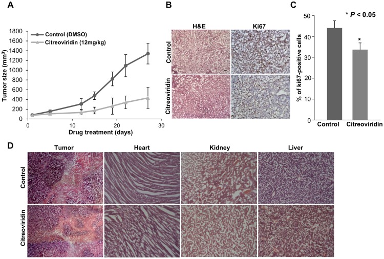
Figure 1. Tumor growth and cell proliferation analysis in the CL1-0 xenograft model.
(A) Tumor regression in a xenograft model. The tumor volume was decreased after treatment with citreoviridin. 5×10⁶ CL1-0 cells were implanted subcutaneously in SCID mice and the abdominal injection of citreoviridin was performed after tumor size reached 100 mm3. (B) The histology (left, H&E, 100×) and Ki67 staining (right, Ki67, 100×) within the same area of tumor tissues. (C) The percentage of proliferating cells in tumor sections using Ki67-immunohistochemistry. Ki67 staining showed a lower percentage of proliferating cells in citreoviridin-treated tumors. (D) Histological analysis of tumor tissues and mice organs. The histology of tumor and organ tissue sections was analyzed by H&E staining (tumor sections, 40×; organ sections, 100×). No obvious histological damages were observed in citreoviridin-treated organ sections, including the heart, kidney and liver. All the staining was performed in 10 µm cryostat sections. H&E, hematoxylin and eosin.

