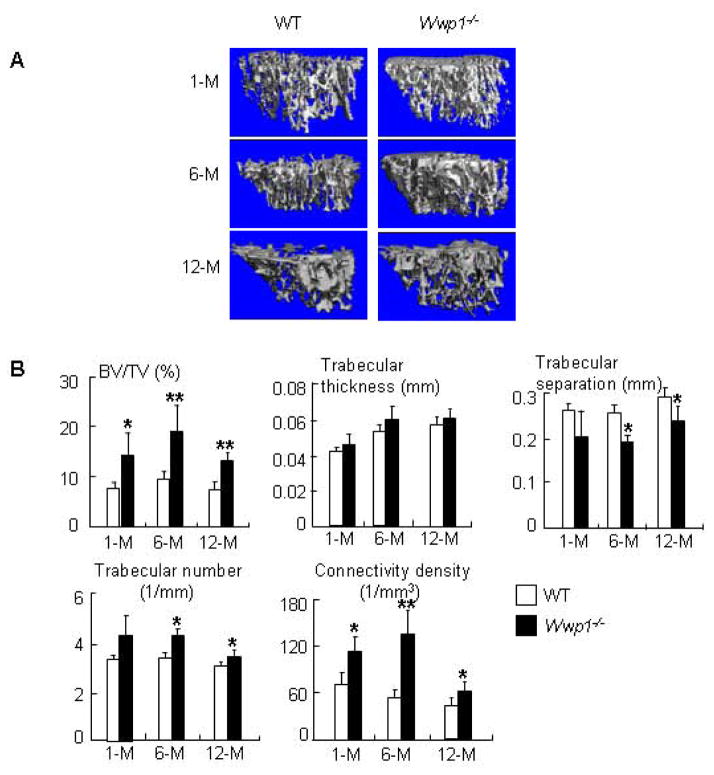
Figure 2. Increased trabecular bone volume in Wwp1−/− mice.
Femurs isolated from 1-month-, 6-month- and 12-month-old Wwp1−/− mice and WT littermates were subjected to micro-CT analysis. (A) Representative 3D reconstructed images show significantly increased trabecular structure parameters in bones of Wwp1−/− mice. (B) Trabecular bone parameters from microCT analysis. Values are the means ± SD of 5 mice. *, p<0.05 vs data from WT mice.

