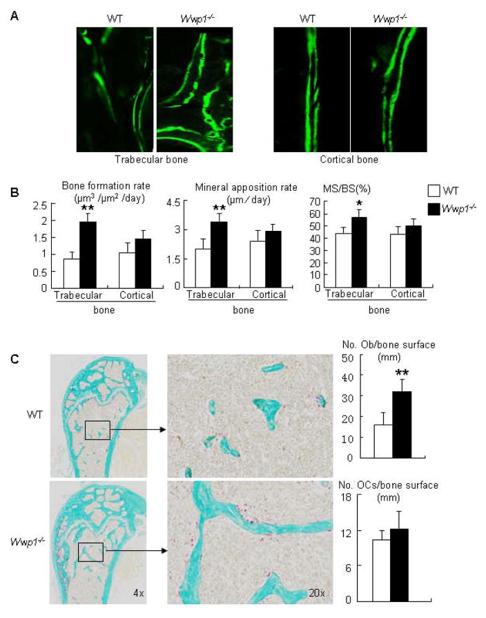
Figure 3. Increased bone formation in Wwp1−/− mice.
Six-month-old Wwp1−/− mice and WT littermates were labeled with calcein, and femurs were subjected to histologic examination. (A) Representative images of calcein-labeled trabecular and cortical bone. (B) Histomorphometric analyses of calcein-labeled sections. (C) Right panels: representative TRAP-stained sections. Left panels: histomorphometric analyses of TRAP-stained sections. Values are the means ± SD of 5 mice. **, p<0.01 vs data from WT mice.

