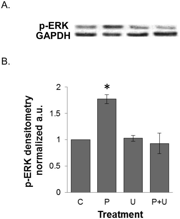
Figure 9.
Western blot analysis and quantitative densitometry of phosphorylated ERK expression in HT-29 cells treated with PPLGM ± a MEK inhibitor. HT-29 cells were treated with the vehicle (C), 10 μM PPLGM alone for 15 minutes (P), 10 μM U0126 alone for 1 hour (U), or pre-treated with 10 μM U0216 for 1 hour followed by treatment with 10 μM PPLGM for 15 minutes (P+U). A) The western blot shows that PPLGM alone enhanced p-ERK expression while U0126 abrogated this effect. B) Densitometry shows the results of PPLGM and/or U0126 treatment on p-ERK expression normalized to GAPDH in three replicate experiments ± standard deviation, where * denotes statistical significance (p<0.05).

