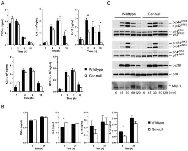
Figure 4.
LPS responses of Gsr-deficient mice and macrophages. A. Serum cytokine and chemokine levels in wildtype and Gsr-deficient mice. Mice were challenged with LPS i.p. (15 mg/g body weight) and euthanized at the indicated time-points. Blood was collected by cardiac puncture, and serum cytokines were measured by ELISA. Values in the graphs represent mean ± SEM of 5–10 independent experiments. B. Cytokines produced by thioglycollate-elicited peritoneal macrophages. Macrophages were stimulated with LPS (100 ng/ml) for 8 or 24 h, and cytokines in the medium were assessed by ELISA. *, p < 0.05 (Student's t-test), compared between the wildtype and Gsr-deficient groups. Values in the graphs represent mean ± SEM of at least 3 independent experiments. C. The activation of MAPKs and Mkp-1 induction in LPS-stimulated peritoneal macrophages. Macrophages were harvested from thioglycollate-stimulated mice, and stimulated with LPS (100 ng/ml). The activation of MAPKs and the induction of Mkp-1 were assessed by Western blot analysis using antibodies against phospho-MAPKs and Mkp-1. The blots were stripped and blotted with antibodies against total MAPKs. Data shown are representative results.

