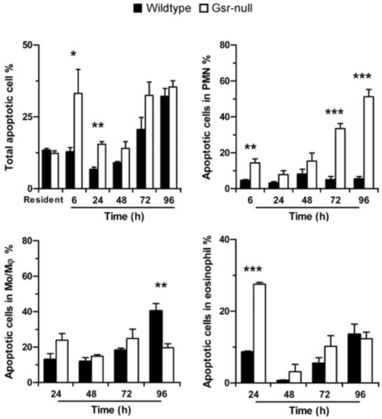
Figure 6.
Increased phagocyte apoptosis in the peritoneal cavity of Gsr-deficient mice after thioglycollate stimulation. Mice were treated as described for Figure 5, and peritoneal leukocytes were harvested by lavage. The percentages of apoptotic cells in total PEC and in distinct leukocyte populations were quantified by flow cytometry after staining with annexin V and PI. Cells that were positive for annexin V were counted as apoptotic cells. Data represent mean ± SEM from at least 3 independent experiments. *, p < 0.05; **, p < 0.01; ***, p < 0.001 (Student's t-test), compared between the two different groups at the same time-points.

