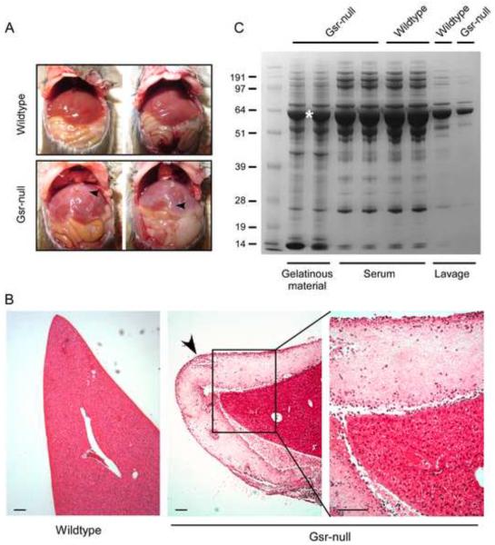
Figure 7.
Demonstration and characterization of protein aggregation coating the internal organs of Gsr-deficient mice after thioglycollate stimulation. Female wildtype and Gsr-deficient mice were stimulated with thioglycollate medium (3%, 2 ml, i.p.) and euthanized 48 h later. The peritoneal cavities were opened for photography, or collection of the coating on the livers. Livers were excised from the animals, fixed, and liver sections were stained with H&E. A. Photographs of the internal organs of wildtype and Gsr-deficient mice. Note the whitish gelatinous coating materials on the liver of Gsr-deficient mice (indicated by arrows). B. H&E staining of the liver sections. Note the layer of fibrous coating on the surface of the liver of the Gsr-deficient mice marked by an arrow. Scale bar represents 100 μm. C. Coomassie blue staining of proteins in the gelatinous material, serum, and lavage samples. Mice were euthanized to collect blood by cardiac puncture, or peritoneal fluids by lavage with PBS. Additional mice were euthanized to collect the gelatinous coating materials on the liver of Gsr-deficient mice. The lavage fluids were centrifuged and supernatants were separated on NuPAGE gel together with serum and the gelatinous coating collected from the surface of the livers of thioglycollate-stimulated Gsr-deficient mice. The band marked with an asterisk was subjected to mass spectrometry analysis.

