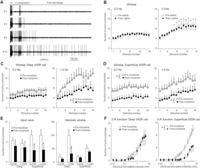
Fig. 1. Spinal administration of morphine induces different inhibitory effects on deep and superficial WDR neurons.

(A) Analog recordings of action potential (AP) responses of a WDR neuron to the first, fourth, eighth, and twelfth stimuli of a train of windup-inducing electrical stimuli (1.0 Hz, supra-C threshold, 2.0 ms). The WDR neuron displayed a progressive increase in the number of APs in the C-component (windup) and after-discharges. The A-component and C-component were divided by the early (0–40 ms) and late (40–250 ms) latencies to a single intracutaneous electrical stimulus. (B) The number of APs in the C-component of WDR neurons (n=8, 5 deep, 3 superficial) in response to repetitive electrical stimulation (0.2 Hz and 1.0 Hz, 16 pulses, supra-C threshold, 2.0 ms) was not changed by topical spinal application of saline. The C-component responses in (C) deep (350–700 μm, n=8) and (D) superficial (<350 μm, n=11) WDR neurons to 0.2 Hz and 1.0 Hz electrical stimuli were decreased 15–30 min after spinal superfusion of morphine (0.5 mM, 30 μl). (E) The input (number of C-component responses evoked by the first stimulus of the train) and absolute windup values before and after morphine treatment in superficial and deep WDR neurons. Absolute windup = (total number of APs evoked by the train) – (16 × input). (F) Stimulus-response (S-R) functions of the steady C-component response to graded electrical stimuli (0.01–5.0 mA, 2.0 ms) were significantly depressed in superficial, but not deep, WDR neurons 15–30 min after morphine treatment. Data are expressed as mean ± SEM. * p<0.05 versus input, # p<0.05 versus pre-drug baseline.
