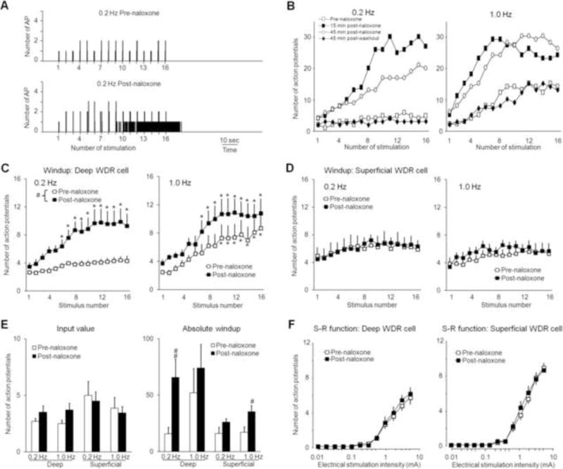
Fig. 2. Spinal administration of naloxone facilitates the development of windup in response to 0.2 Hz stimulation in deep WDR neurons.

(A) Histograms show that the evoked responses of a WDR neuron to 0.2 Hz intra-cutaneous electrical stimuli (16 pulses, supra-C threshold, 2.0 ms) were markedly increased after naloxone treatment (1 mM, 30 μl). Bin sizes were 10 ms. (B) Representative graphs show the time course by which naloxone facilitates windup in a deep WDR neuron (476 μm) at 0.2 Hz and 1.0 Hz stimulation. The windup curves show the C-component responses in (C) deep (350–700 μm, n=10) and (D) superficial (<350 μm, n=8) WDR neurons to 0.2 and 1.0 Hz electrical stimuli before and 15–30 min after spinal superfusion of naloxone. (E) The input and absolute windup values before and after morphine treatment were plotted for the various frequencies in deep and superficial WDR neurons. (F) Stimulus-response (S-R) functions of C-component in deep and superficial WDR neurons to graded electrical stimuli (0.01–5.0 mA, 2 ms) did not change 15–30 min after naloxone treatment. Data are expressed as mean ± SEM. * p<0.05 versus input, # p<0.05, ## p<0.01 versus pre-drug baseline.
