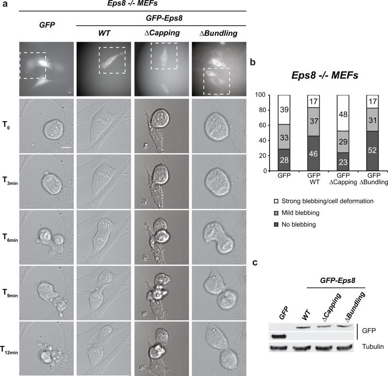
FIGURE 7. MEF Eps8 -/- cells exhibit a post-metaphase membrane blebbing phenotype that can be rescued by re-expression of Eps8 versions with actin capping activity.
(a) Still images of DIC time lapses of mitotic mouse embryo fibroblasts (MEFs). MEFs derived from Eps8 -/- mice5 and infected with either control pBABE-GFP vector (GFP) or pBABE-GFP-Eps8 WT, ΔCapping, or ΔBundling were seeded onto gelatine-coated coverslips at low confluency. The day after seeding, cells were subjected to time-lapse analysis (24 hours, time interval 2 minutes). 30 MEFs passaging through mitosis were recorded and classified per condition. Upper panels: fluorescence images taken immediately before the time-lapse. Lower panels: DIC images of the GFP-positive cells (white dashed line insets in upper panels) followed during mitosis. Bar = 10 μm.
(b) Quantification of the experiment in (a).
(c) Immunoblot analysis of expression levels of GFP-Eps8 versions in infected MEF Eps8 -/- cells. α-tubulin levels serve as loading control.

