FIG. 7.
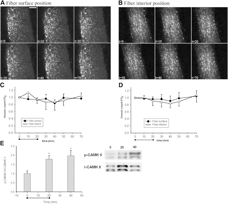
Caffeine-mediated Ca2+/calmodulin-dependent protein kinase II (CAMK II) phosphorylation does not reduce IL-6-EGFP or EYFP-Golgi vesicle abundance. A and B: t = 0 shows confocal images of basal IL-6-EGFP vesicles in the surface (A) or interior (B) part positions of a mouse quadriceps muscle fiber prior to intravenous caffeine infusion. Similar observations were made in fibers from six mice. Bar = 20 μm. Immediately after t = 0, caffeine infusion was initiated for 20 min and confocal images were collected every 10 min during and after infusion. C and D: Image quantification of IL-6-EGFP (C) or EYFP-Golgi (D) vesicles from images taken at the surface position or interior position throughout the time period after i.v. infusion of caffeine. Values are mean ± SE, n = 6. E: Western blot of phosphorylated Ca2+/calmodulin-dependent protein kinase II from muscle either basal, 20, or 40 min after caffeine infusion. Values are mean ± SE, n = 5–6. *P < 0.05.

