FIG. 8.
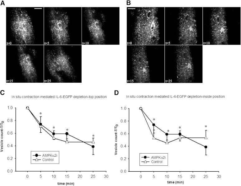
AMPK activation is not essential for IL-6-EGFP vesicle reduction. A and B: t = 0 shows confocal images of basal quadriceps muscle fibers expressing IL-6-EGFP prior to in situ contractions from either a wild-type control (A) or an AMPKα2-inactive transgenic (B) mouse. IL-6-EGFP is localized to vesicle-like structures near the surface position in the muscle fibers. The interior positions are not shown but demonstrate similar vesicle distribution. In situ contractions were elicited for 3 × 5 min periods (t = 5, 10, 15) and then a 10-min period (t = 25), each separated by 90 s of rest. Similar observations were made in fibers from 5–6 mice. Bar = 20 μm. C and D: Image quantification of IL-6-EGFP vesicles from images taken at the surface (C) or interior (D) positions of the muscle fibers in AMPKα-inactive transgenic mice and wild-type control mice following each contraction period. Values are mean ± SE, n = 5–6. *P < 0.05 compared with t = 0.

