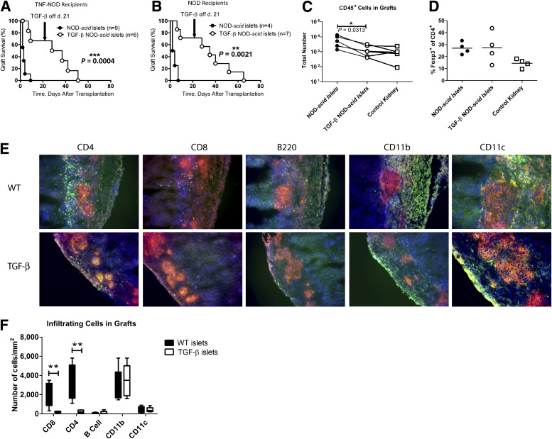
FIG. 1.
Induced expression of TGF-β for the first 21 days after transplantation protects islet grafts from destruction. RIP-TNF NOD mice (A) or WT NOD mice (B) were confirmed to be diabetic and transplanted with an islet graft from either NOD-scid (●) or TGF-β–NOD-scid mice (○). The difference between the groups was plotted using a Kaplan-Meier plot, and differences between groups were determined using the log-rank test. Absolute numbers of cells infiltrating the grafts of RIP-TNF NOD mice were assessed by flow cytometry 5 days after transplantation (C). Lines connect grafts performed on the same occasion, and the Wilcoxon signed rank test was used to assess differences between groups. The percentage of Foxp3+ cells in the CD4+ T-cell gate in these cells was assessed (D). Sections of the grafts from RIP-TNF NOD mice were stained for insulin (red) and the indicated immune cell (green). DAPI was used as a nuclear stain (blue) (E). Three fields of vision were assessed for at least five mice from each group, and the numbers of infiltrating cells of the indicated subtypes were determined (F). Differences between groups were determined using the Mann-Whitney test (P = 0.0079 for CD8+ T cells and P = 0.0079 for CD4+ T cells). *P ≤ 0.05, **P ≤ 0.01, ***P ≤ 0.001.

