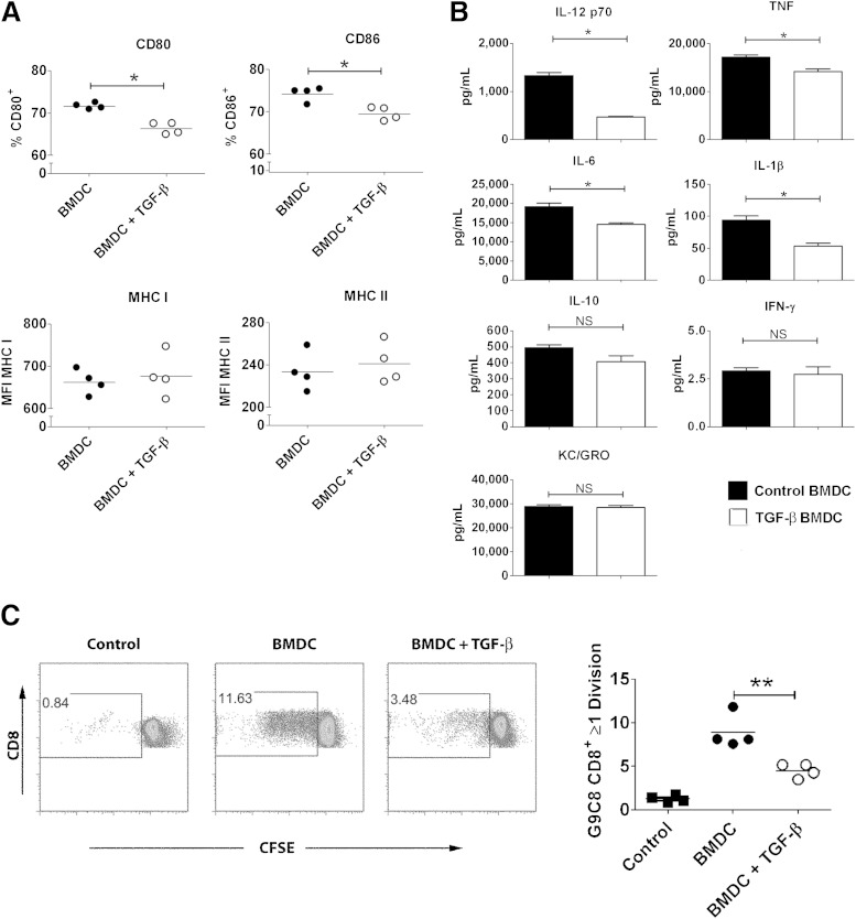
FIG. 5.
Exposure to TGF-β reduced expression of costimulatory molecules on BMDCs and reduced their capacity to stimulate T cells. NOD BMDCs were activated with 100 ng/mL LPS after preconditioning overnight with 5 ng/mL TGF-β. Expressions of costimulatory molecules CD80 and CD86 as well as MHC class I and class II were assessed using flow cytometry (A), and production of cytokines in supernatants was determined using seven-plex analysis (B). NOD BMDCs were activated with LPS after preconditioning overnight with 5 ng/mL TGF-β, pulsed with insulin β-chain peptide 15-23 (ins15-23), and cultured for 72 h with CFSE-labeled insB15-23–specific CD8+ T cells from the G9 TCR transgenic mouse. Proliferation was assessed by flow cytometry. Filled squares, proliferation without DC; filled circles, proliferation with control BMDC; open circles, proliferation with TGF-β–exposed BMDC (C). Differences in surface markers and cytokine expression between groups were determined using the Mann-Whitney test, whereas differences in proliferation were assessed using the Student t test. Results show technical replicates and are representative of at least three independent experiments. NS, not significant. *P ≤ 0.05, **P ≤ 0.01.

