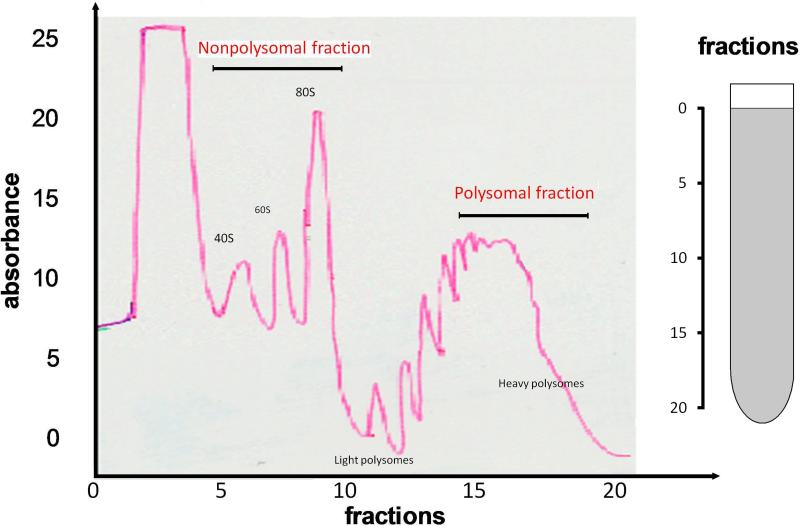
Fig.3. Realtime OD detection of the polysome fractions.
Fractions were monitored using an ISCO UA-6 UV detector. The positions of 80S ribosomes, light polysomes, and heavy polysomes, in the gradients are labeled. Two fractions, i.e. nonpolysomal fraction (corresponds to Fraction 7-10) and polysomal fraction (corresponds to Fraction 15-18), used in this study were indicated with arrows. X-axis: the fraction number (from Fraction 1 to Fraction 24); Y-axis: the UV absorbance. Increases in polysome size by a single ribosome are indicated by secondary peaks in the up-slope of the broad polysome peak.

