Fig. 2.

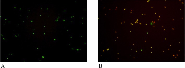
Fluorescenct micrographs of spores of T. rubrum. A, Control spores showing green fluorescent, live cells; B, Limonene treated spores at 0.5% v/v showing orange fluorescent, dead cells.

Fluorescenct micrographs of spores of T. rubrum. A, Control spores showing green fluorescent, live cells; B, Limonene treated spores at 0.5% v/v showing orange fluorescent, dead cells.