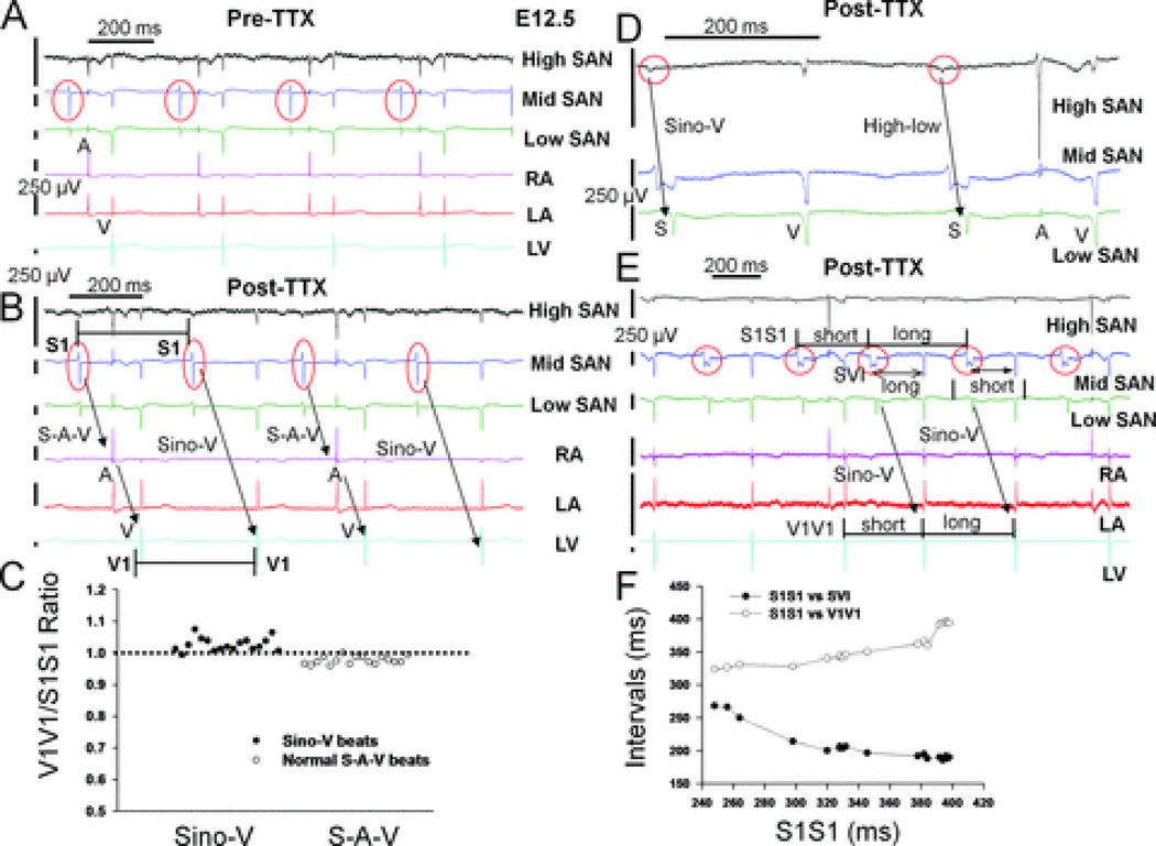
Figure 5. Sino-ventricular linking during Sino-V conduction and evidence for internodal conducting pathways.
A: Raw MEA tracings from an E12.5 heart before and B: after TTX application. Electrical recordings from three consecutive electrodes around the SAN region and at a cranial-to-caudal sequence were named high, mid, or low SAN tracings in this figure (see also Supplements Fig. 3). TTX led to a stable pattern of alternating (2:1) normal sino-atrial-ventricular (S-A-V) (first and third beats) and Sino-V conduction sequences (second and fourth beats). C: During 12 sec recordings of stable 2:1 Sino-V conduction rhythm, the ratio of ventricular intervals (V1V1) to preceding sinus cycle length (S1S1) is close to 1 for all normal (S-A-V) and Sino-V conducted beats, indicating SAN-V linking. D: For another E12.5 heart with sinus arrhythmias, the high-to-low conducting sequence remains the same between normal and Sino-V conducted beats, as well as E, the sino-ventricular interval (SVI) during Sino-V conduction is longer after a short preceding S1S1 interval. F: Using all Sino-V conducted beats from a 30 sec recording period shown in E, a plot of V1V1 intervals and SVIs versus preceding S1S1 intervals showed a progressive increase in SVIs when S1S1 intervals were shorter than 320 ms, which is a classic decremental conduction property of AVN and supports an inter-nodal pathway that mediates Sino-V conduction with AVN as the relay. Red circles indicate sinus signals.

