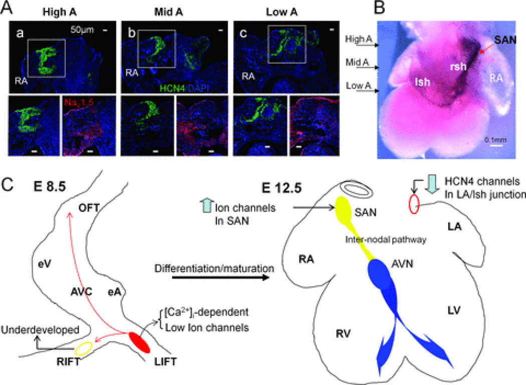
Figure 6. Immunostaining and In situ hybridization of HCN4 channels subunits revealed a potential internodal pathway at E12.5.
A: Consecutive sections of E12.5 atria from high to low levels were obtained for immunostaining (only three levels of sections are shown here). Immunostainings of high atria (High A) at SAN level, middle atria (Mid A) and low atrial (Low A) close to AVN level are shown. Magnified views at each level showed a pathway throughout these atrial levels with high HCN4 staining (lower left) and low NaV1.5 expression (lower right) at the RA-right sinus horn (rsh) junction. B: In situ hybridization (dorsal view) is used to show a continuous HCN4 expression pathway in the RA with arrows indicating comparable levels shown in A. C: Schematic summary of location and mechanistic switch of dominant automaticity during cardiac differentiation. Left panel, in E8.5 hearts, intracellular Ca2+-driven mechanisms dominate control of the LIFT pacemaker site (red region) with small contributions of sarcolemmal ion channels to automaticity and an underdeveloped RIFT pacemaker site (yellow circle). Right panel, in E12.5 hearts, ion channels up-regulate (an arrow pointing up) in the right-sided SAN (filled yellow circle) and control the dominant rhythm with concomitant down-regulation (an arrow pointing down) of HCN4 channels in the comparable region at LA/ left sinus horn junction (red circle). An internodal conduction pathway (a yellow tract) could be demonstrated functionally in E12.5 hearts. The normal AVN-bundle branch-Purkinje system is labeled with blue color.

