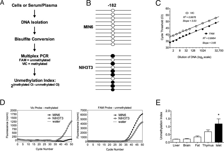
Figure 1.
Unmethylated and Methylated PPI DNA Is Detectable in Mouse Cell Lines and Tissue. A, DNA was extracted from mouse cell lines, tissue, serum or plasma, bisulfite treated, and multiplex PCR was performed. B, Methylation pattern of the −182 CpG site of the PPI promoter with each line representing an individual clone. Open circle, unmethylated; closed circle, methylated. C, Real-time cycle Ct values determining linearity of PCR amplification. D, Quantitative real-time PCR of the methylated product using the VIC-labeled probe and the unmethylated product using the FAM-labeled probe in MIN6, NIH3T3, and water control samples. The gray line indicates the threshold. E, Unmethylation index in mouse liver, brain, fat, thymus, and islet tissues. n = 3–5; *, P < .05.

