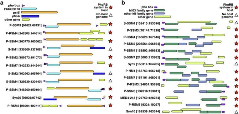
Figure 3.
Predicted pho boxes immediately upstream of (a) PhCOG173 and/or pstS and (b) the hli03 gene cluster in cyanomyovirus genomes. Phage genome names and the genomic indices of the displayed region are indicated. Putative pho box motifs are shown as purple arrows. The genomic region in (a) is larger than the region in (b) and the pho box motif and genes are scaled in size accordingly. Red stars indicate that the host strain on which the phage was isolated contained the PhoBR two-component phosphate sensing system; white triangles indicate that the host genome is not currently available. The PhCOG173 (cyan), pstS (orange), phoA (blue), hli03 (dark green) and other hli genes (light blue) are highlighted with specific colors; all other genes are shown in light green.

