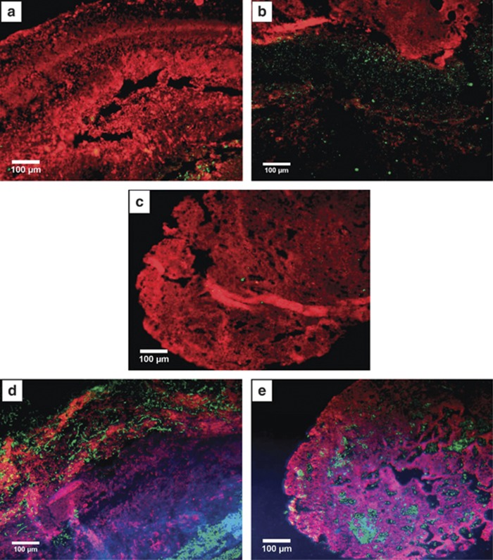
Figure 3.
CARD FISH pictures of a cross section of the snottite. (a) In the upper part (Spot 1, Figure 1A) of the snottite bacteria (EUB338, Alexa 546 in red) can be seen throughout the whole cross section while the archaeal signal (ARCH915, Alexa 488 in green) is only in the middle part, which is most likely fully anoxic. (b) Inner area of the upper part of the biofilm (Spot 1, Figure 1A). Archaea (ARCH915, Alexa 488 in green) are found in high abundance. (c) Bacteria (EUB338, Alexa 546 in red) and archaea (ARCH915, Alexa 488 in green) in the lower part of the biofilm (Spot 4, Figure 1A). The image shows that the whole biofilm is populated by bacteria but almost no archaea-derived signal is detectable. (d) Image of the upper part (Spot 1, Figure 1A) of the snottite. The Acidithiobacillus species (THIO1, Alex 488 in green) are in the very outer and in the inner part while we see Leptospirillum ferrooxidans (LEP636, Alexa 546 in red) in a broad outer area. DAPI (4,6-diamidino-2-phenylindole) in blue shows all cells. (e) In the lower part of the snottite (Spot 4, Figure 1A) where no anoxic zone was detectable (Spot 4, Figure 1A), an unspecific distribution of the Leptospirillum (LEP636, Alexa 546 in red) and Acidithiobacillus (THIO1, Alex 488 in green) species is found. DAPI stain shows all organisms in blue.

