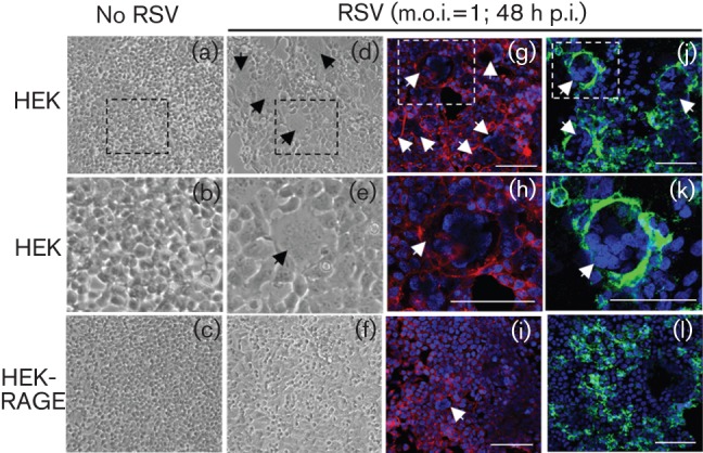
Fig. 3.

RAGE inhibits RSV-induced syncytium formation. HEK and HEK-RAGE cell cultures were infected with live RSV (m.o.i. = 1) for 48 h as indicated and photographed. Light micrograghs (a–f) were taken at 10× magnification and syncytia are indicated with black arrows; the boxed regions in (a) and (d) are enlarged in (b) and (e), respectively. Confocal images (g–l) were taken at 40× magnification and syncytia are indicated with white arrows. Cells were visualized with a membrane stain (red; g–i), Alexa Fluor 488-labelled anti-F protein antibody (green) (j–l) and Hoechst 34580 nuclei staining (blue) (g–l). The boxes in (g) and (j) are enlarged to show increased detail in (h) and (k), respectively. Bars, 100 µM (g–l).
