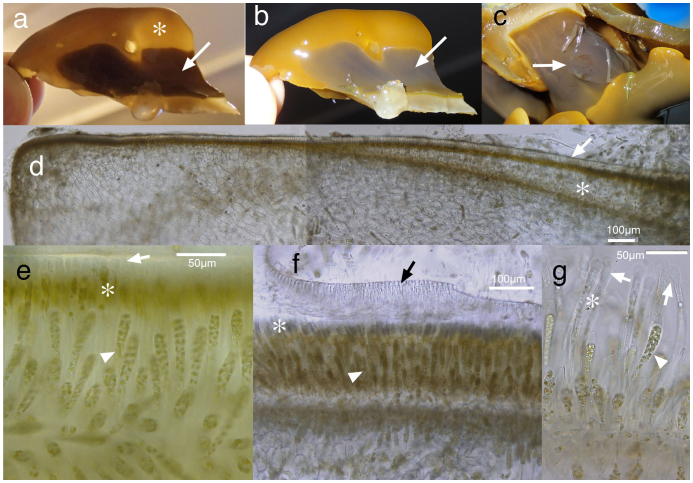
Figure 3. Morphology and anatomy of the basal system of Aureophycus aleuticus (St. George Is., 22–24 October 2012).
(a, b), sorus under transmitted (a) and strong epi-illumination showing iridescence of sorus (b). Note the difference between vegetative portion (asterisk) and sorus (arrow). (c), fertile sorus showing partly detached cuticle (arrow). (d), cross section of basal system (discoid holdfast) showing the development of sorus. Note that the space between cuticle (arrow) and original peripheral layer becomes thicker toward the right side of the section (asterisk) by the development of paraphyses; (e, f), detachment of cuticle (arrow) by the expansion of mucilaginous caps (appendages) at the tips of paraphyses (asterisk). Arrowhead shows unilocular zoidangia. (g), unilocular zoidangium (arrowhead), paraphysis (asterisk) and mucilaginous cap (arrows). [Photographs by H. Kawai].

