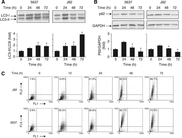
Figure 2.
Pazopanib triggers the autophagic process in BC cells. (A and B) Lysates from 5637 and J82 cell lines, untreated or treated with 20 μM pazopanib at different times, were separated on 14% or 10% SDS–PAGE and probed with anti-LC3 or anti-p62 and anti-GAPDH Abs, respectively. Densitometric analysis, expressed as the mean±s.d., was performed by comparing treated with untreated cells, *P<0.01. (C) The percentage of bright red fluorescence (FL3) cells was evaluated by AO staining and FACS analysis in 5637 and J82 cells untreated or 20 μM pazopanib treated at different times. Dot plots represent one out of three separate experiments.

