
Figure 5.
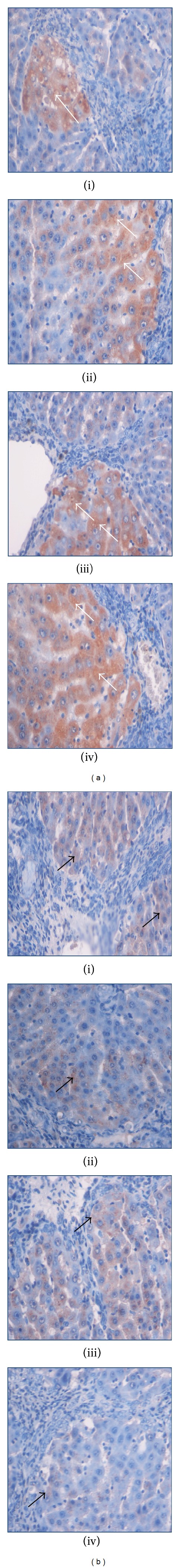
Immunohistochemistry staining of (a) Bax and (b) Bcl-2 of representative livers sampled from rats in different experimental groups. Less apoptosis indicated by (ia) few Bax-positive hepatocytes (white arrow) and (ib) more Bcl-2-positive hepatocytes (black arrow) in liver tissues from a cirrhosis rat group. (iia) High numbers of Bax-positive hepatocytes (White arrow) and (iib) very less Bcl-2-positive hepatocytes (black arrow) in the liver from a hepatoprotected rat treated with Silymarin. Moderate apoptosis as indicated by (iiia) moderate Bax staining (white arrow) and (iiib) moderate Bcl-2 staining (black arrow) indicating moderate apoptosis in the liver from the rats treated with low dose BR extract. (iva) High numbers of Bax-positive cells (white arrow) with severe apoptosis and (ivb) very less Bcl-2-positive hepatocytes (black arrow) were observed in the liver of the rats treated with high dose BR extract (original magnification ×40).