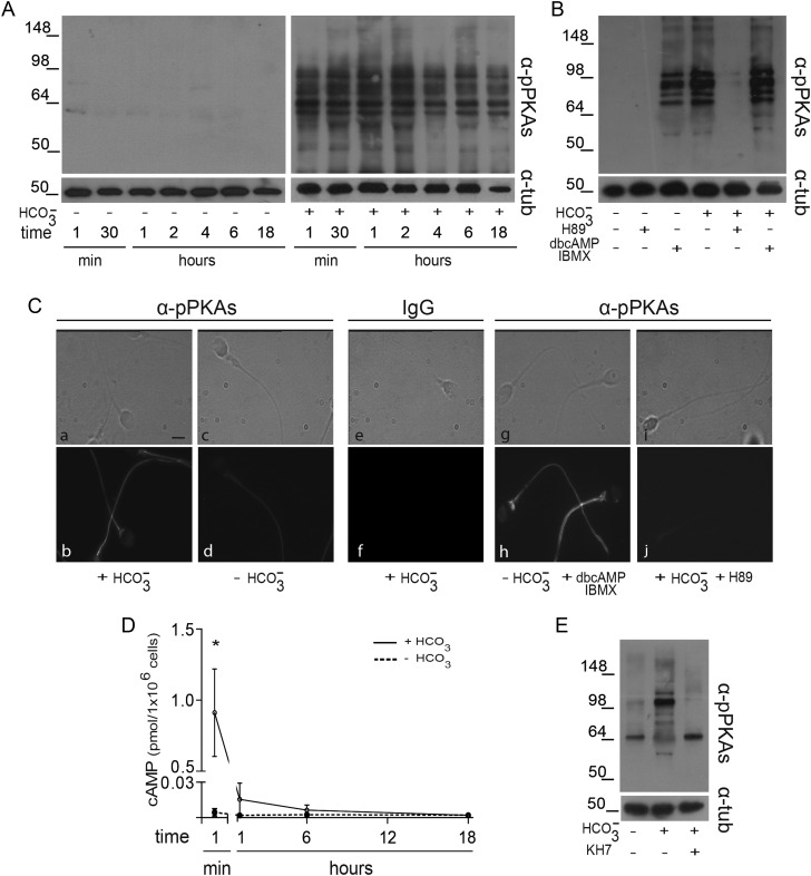
Figure 1.
Evaluation of PKA activity during human sperm capacitation. (A) Sperm were incubated in media with (right panel) or without (left panel)  for different time periods (1–18 h). Aliquots were removed at different intervals and sperm proteins were analyzed for PKA substrate phosphorylation by western blotting using α-pPKAs as the first antibody. β-tubulin was used as control of loading (n = 8). (B) Sperm were incubated for 18 h in media with or without
  and containing either H89 (30 µM) or dbcAMP/IBMX (5 mM/0.2 mM). Protein extracts were analyzed for PKA substrate phosphorylation by western blotting (n = 5). (C) Phase-contrast (upper) and fluorescent (bottom) images of sperm incubated for 1 min in media with (a, b, e, f) or without (c, d)
  , in media without
  but containing dbcAMP and IBMX (g, h), or in complete media with H89 (i, j). The IIF was performed using α-pPKAs (a–d, g–j) or purified IgG as first antibodies (e, f) (n = 4). The scale bar is 5 μm. The same images were observed for sperm incubated for 1 or 18 h. (D) Cell extracts from sperm incubated in media with (solid line) or without (dotted line)
  for different times (1 min to 18 h) were analyzed for total cAMP intracellular levels. Values represent the mean ± SEM of four independent experiments. *P < 0.05. (E) Sperm were incubated in media with or without
  in the absence or presence of KH7 (75 µM), and aliquots were removed at 1-min incubation and analyzed for PKA substrate phosphorylation by western blotting (n = 3).

