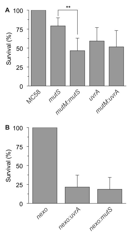
Figure 5.
Contribution of the MMR and NER to the repair of oxidative DNA damage.
(A) Strains (indicated below each bar) were incubated in the presence of 0.2 mM H2O2 and 15 mM paraquat or PBS at room temperature for 30 min and then plated to solid media. Percentage killing was calculated by comparing the number of CFU recovered in the presence or absence of oxidizing agents, and are given as the percentage survival relative to MC58. Error bars show the SD of assays performed in triplicate (** indicates p < 0.01, Students t test). (B) Strains (indicated below each bar) were incubated in the presence of 0.4 mM H2O2 or PBS at room temperature for 30 min and then plated to solid media. Percentage survival was calculated by comparing the number of CFU recovered in the presence and the absence of oxidizing agents, and are given as the percentage survival relative to nexo mutant.

