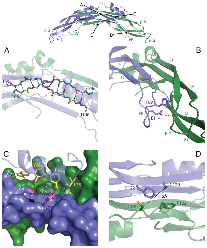
Figure 2. Intermolecular contacts of PRDC dimers.
Top, PRDC dimer side view colored as in Fig 1A. Letters correspond to the general location of close-up views depicted in the panels below. (A) Close-up view of the anti-parallel interaction of β2 of each PRDC monomer. Backbone atoms are shown as sticks and mainchain H-bonds between strands are indicated. (B) Interactions of the wrist (blue) and F1 (green) regions on opposing monomers. View is down the long axis to highlight the 4-stranded β-sheet generated from merging the two monomers. (C) Interaction of the N-terminal helix (transparent) from Chain B (blue) with each monomer, representing the intra-chain (purple) and inter-chain (yellow) residues as sticks at the hydrophobic interface. (D) Zoomed in view showing the spatial relationship of C120 at the dimer interface, indicating the absence of a disulfide bond. See also Figure S2.

