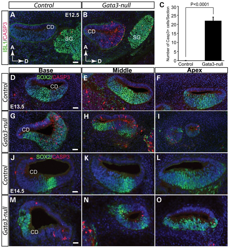
Figure 5.
Elevated cell death in the cochlear duct of Gata3-null mice. (A and B) At E12.5, no CASP3+ apoptotic cells are detected in wild-type mice (A). However, cell death is greatly increased in the cochlear duct of Gata3-null mice (B). (C) Quantification of apoptotic cells (CASP3+/section) in the cochlear duct (P < 0.0001, Fisher's exact test, n = 3 for Gata3-nulls and n = 3 for controls). (D–I) Cell apoptosis analysis reveals dramatically elevated cell death in the cochlear duct of Gata3-null mice at E13.5. By E14.5, the structure of the cochlear duct is disrupted in Gata3-null mice due to cell death (J–O). Compared with wild-type mice, the cochlear duct is much smaller in Gata3-null mice. Data are shown as the mean ± SEM. CD, cochlear duct; SG; spiral ganglion neurons; D, dorsal; A, anterior. Scale bar, 50 µm.

