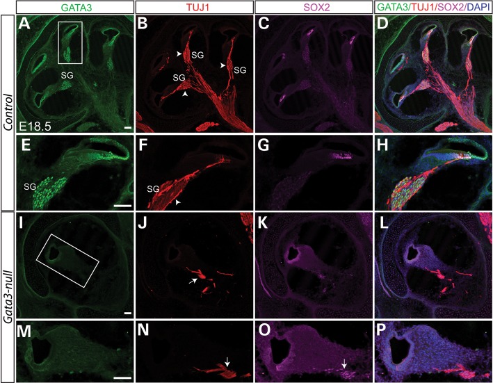
Figure 6.
Depletion of SGNs in Gata3-null mice. (A–D) Triple-immunolabeling shows that GATA3 is highly expressed in the SOX2+ supporting cells and the TUJ1+ SGNs. The peripheral processes of SGNs make synaptic contact with the hair cells while the SGN axons are bundled together to form the auditory portion of eighth cranial nerve (B–D). Contrasting to the numerous SGNs in wild-type mice (arrowhead in B and F), the SGNs are nearly depleted (I–P) and very limited fibers remained (arrow in J and N) in Gata3-null mice. (E–H and M–P) High magnification of boxed regions in (A) and (I). Scale bar, 100 µm.

