
Figure 7.
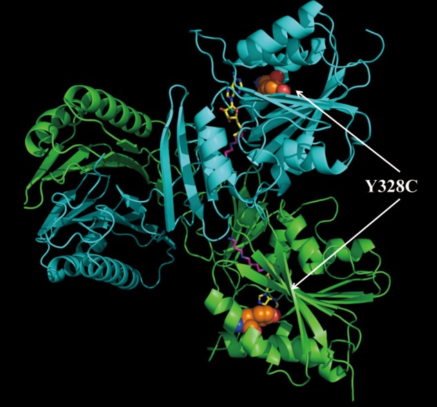
The 3D structure of the SMS dimer. The green one is the C chain, while the cyan one is the D chain. The disease-causing mutation site is shown with colored balls (orange, carbon atom; red, oxygen atom; and blue, nitrogen atom); SPD and MTA are represented with magenta stick and yellow stick, respectively.