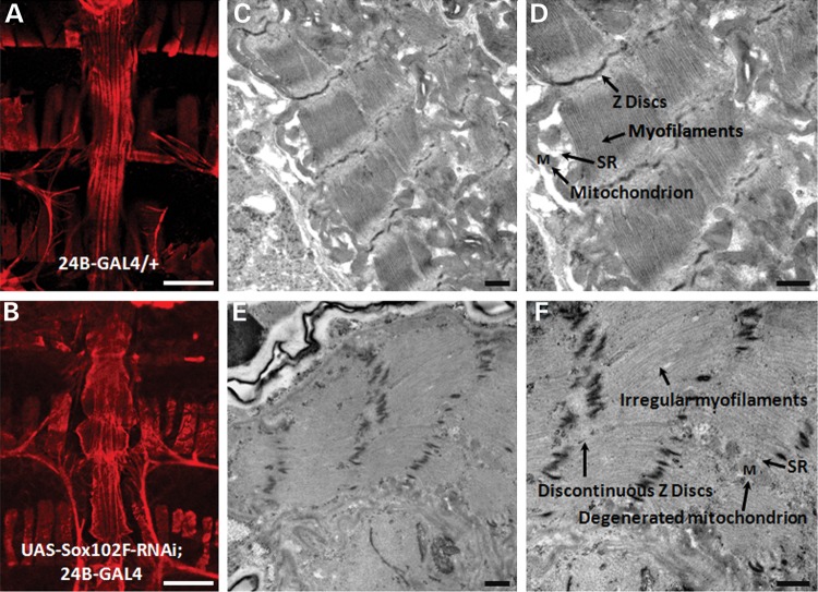
Figure 4.
Whole heart and cardiac ultrastructure analysis. (A and B) Micrographs of the whole adult heart by F-actin immuno-fluorescent staining. (A) Control fly (24B-GAL4/+) showed a normal cardiac tube. (B) Silencing of Sox102F in the heart (UAS-Sox102F-RNAi; 24B-GAL4) resulted in an enlarged and irregular cardiac tube and loss of myofibril structure. (C–F) Ultrastructure of adult heart longitudinal sections between A1 and A3 segments by TEM. (C and D) Control fly showed normal myofibril structure. (E and F) Silencing of Sox102F in the heart led to irregular myofilament arrays, discontinuous Z discs, irregular and smaller SR and degenerative mitochondria. Myofilaments, Z discs, SR and mitochondrion were labeled and pointed with arrows. Magnification: (A and B) 10×, scale bars: 100 µm; (C and E) 20 000×; (D and F) 30 000×, scale bars: 500 nm.

