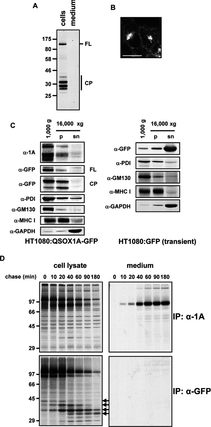
Figure 4. Cleavage products of QSOX1A–GFP.
(A) Western blot showing the full-length GFP-tagged form (FL) of QSOX1A and cleavage products (CP). The cell lysate and serum-free culture medium were harvested following 3 h of incubation in serum-free medium. The GFP-containing fragments were isolated using GFP-Trap®_A and identified by an anti-GFP Western blot. Molecular masses of marker proteins are indicated (in kDa). (B) Live-cell image of QSOX1A–GFP-expressing HT1080 cells by confocal microscopy. The GFP fluorescence is detected in the Golgi, the cell membranes and small vesicles. Scale bar, 20 μm. (C) Western blot analysis of the fractionation of cellular components by differential centrifugation. Cell lysates were homogenized and followed by two consecutive centrifugation steps at 1000 g and 16000 g. The pellet of the first step contains nuclear membranes, the ER and the Golgi. The pellet (p) of the 16000 g centrifugation step enriches the cell membranes and the supernatant (sn) represents the cytoplasm. Nitrocellulose membranes were probed for cleaved and full-length forms of QSOX1A–GFP (α-1A), the GFP-tagged full-length form (α-GFP, FL) and cleavage products (α-GFP, CP), the ER marker PDI (α-PDI), the Golgi marker GM130 (α-GM130), a marker for the cell membrane (α-MHC I) and a cytoplasmic marker (α-GAPDH). Cell lysates were prepared from HT1080 cells stably expressing QSOX1A–GFP (HT1080:QSOX1A-GFP) and from HT1080 cells transiently transfected with soluble GFP (HT1080:GFP) respectively. (D) Pulse–chase experiments illustrating the cleavage of QSOX1A–GFP. The samples were radiolabelled for 30 min and secretion followed by harvesting cells and medium at the indicated time points. Samples were immunoisolated by using either the anti-QSOX1A antibody bound to Protein A–Sepharose or GFP-Trap®_A. The samples were separated by SDS/PAGE and gels were exposed to Kodak imaging film. Cleavage products are marked by arrows. Molecular masses of marker proteins are indicated (in kDa). IP, immunoprecipitation.

