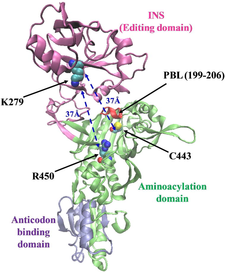
Figure 1.

Ribbon representation of the 3-dimensional structural model of the monomeric form of Ec ProRS. The homology model was derived from the X-ray crystal structure of Ef ProRS (15). The Cα – Cα distances between the starting residues (C443/R450) and the end residue (K279) are shown.
