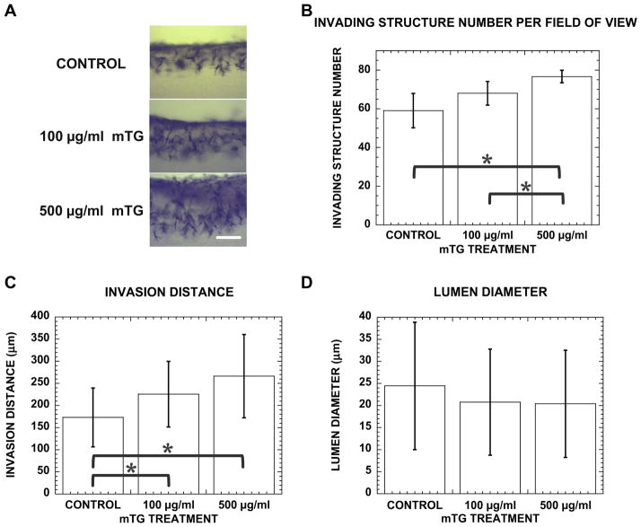
Fig. 6.
Stiffer matrix promotes angiogenic invasion with more structures invading deeper into collagen gel. (A) Cross-sectional images of control, 100 and 500 μgml−1 mTG-treated cultures stained with toluidine blue at 48 h. Scale bar is 250 μm. Quantification of (B) invading structure number, (C) invasion distance and (D) lumen diameter in control, 100 and 500 μg ml−1 mTG-treated collagen gels at 48 h. Sample number n = 4 gels in (B), n = 130, 101, 149 structures in (C) and n = 53, 37, 46 structures in (D) for control, 100, 500 μgml−1 mTG-treated gels, respectively. Symbol * represents P < 0.05.

