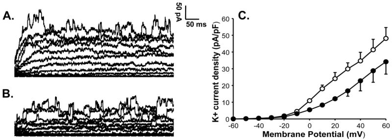
Figure 4.

K+ current of a pulmonary artery VSMC from a control lamb is shown at basal level (A) and after application of 4-AP (B). K+ current shows significant suppression by 3 mM 4-AP indicating the presence of Kv channel activity in a control cell. Summarized data from 4 cells is shown to the right in panel C for K+ current density in the absence (-○-) or presence of 4-AP (-●-) and demonstrates inhibition of the current by 4-AP.
