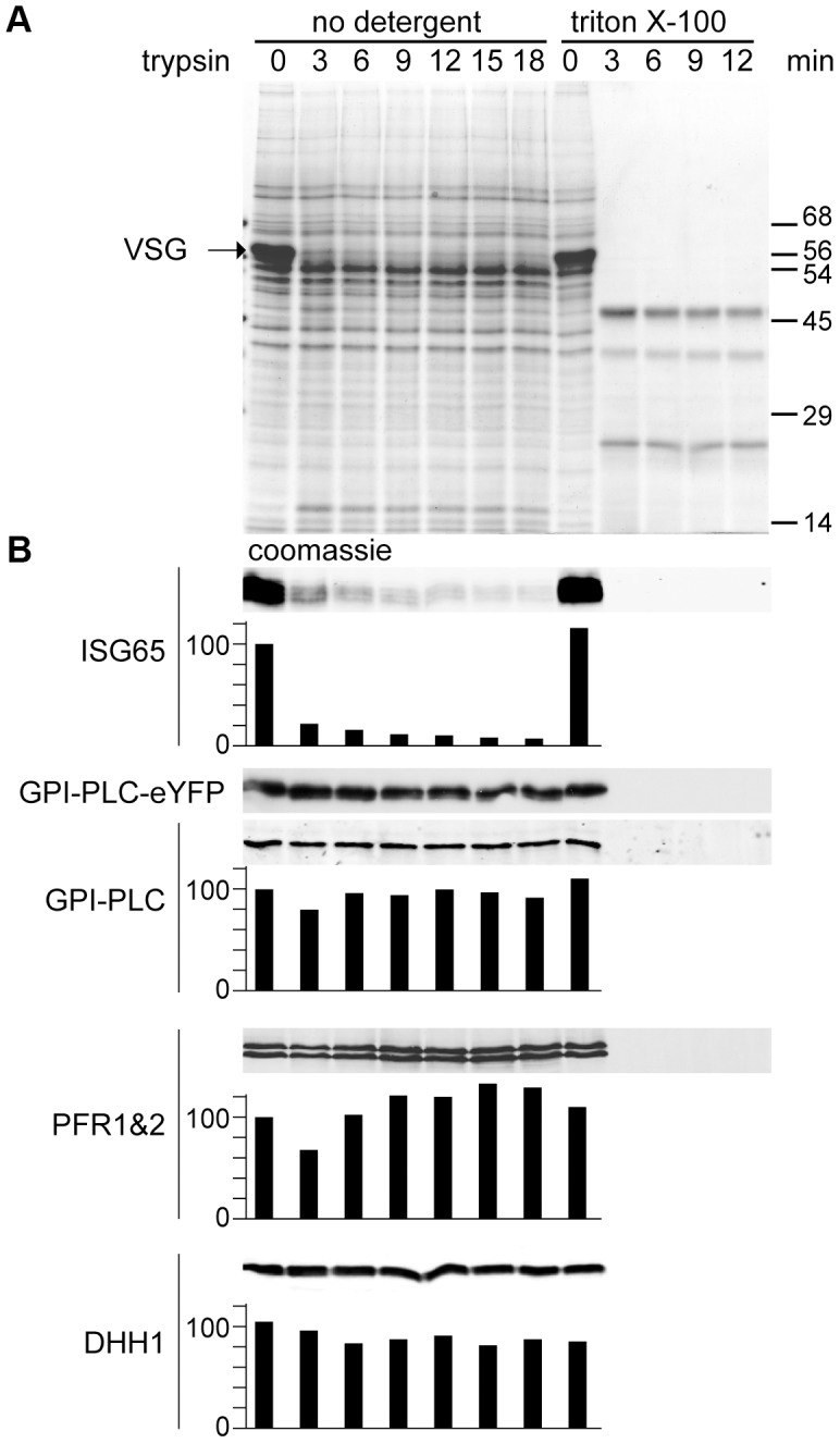
Figure 2. GPI-PLC is not sensitive to trypsin digest in live cells.

A cell line modified to contain one copy of the wild type GPI-PLC gene and one copy modified with a C-terminal eYFP tag (GPI-PLC-eYFP/+) was treated with exogenous trypsin and samples were removed over a time course for analysis by SDS-PAGE and Western blotting. A parallel time course after 0.5% triton X-100 addition was used as a control for trypsin sensitivity. (A) Coomassie stained gel of a trypsin digest time course of GPI-PLC-eYFP/+ cells. Note the almost complete digestion of VSG by 3 minutes. (B) Western blots with relative quantitation below, setting the zero time point at 100. Quantitation used an Odyssey Infrared Imaging System and associated software. However, the expression levels of GPI-PLC-eYFP were too low for reliable detection using fluorescent secondary antibodies and were instead detected by chemiluminescence; this was not quantitated. ISG65 was mostly digested within 3 minutes. GPI-PLC-eYFP and GPI-PLC were stable over the time course but were digested within 3 minutes when detergent was included as were the flagellar proteins PFR1 and 2 and the cytoplasmic protein DHH1. 2×10⁶ cell equivalents were loaded in each track.
