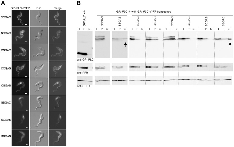
Figure 4. Cysteine mutants mis-localise to the endosome and/or cytoplasm.
(A) Cell lines modified to contain a single copy of the wild type (CCGAC) or cysteine mutant GPI-PLC all modified with a C-terminal eYFP tag were imaged using the native fluorescence of the eYFP tag after being immobilised. Each image is representative of the distribution observed in the majority of cells. Scale bar represents 2 µm. (B) Wild type (GPI-PLC +/+) and the same cell lines as (A) were hypotonically lysed for 5 minutes on ice and the lysate (I) separated into supernatant (S) and pellet (P) fractions and analysed for the distribution of GPI-PLC-eYFP by Western blotting. The detection of PFR is a control for the cytoskeleton present in the pellet and DHH1 for cytoplasmic proteins. The arrows indicate the presence of GPI-PLC-eYFP in the supernatant fraction. 2×10⁶ cell equivalents were loaded in each track and the blots were cut for presentation purposes.

