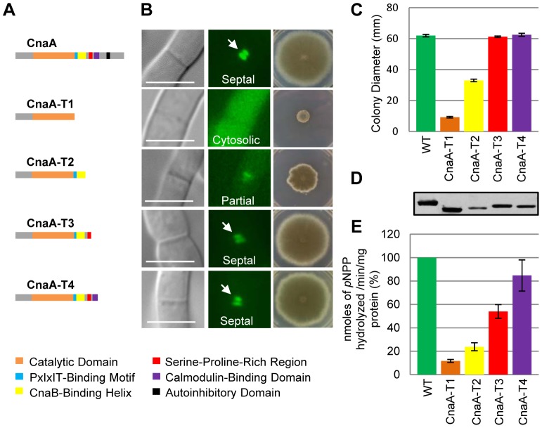
Figure 1. Truncations of A. fumigatus CnaA revealed important domains required for growth and septal localization.
(A) Scheme of cnaA truncations and domain organization. Constructs were expressed with native cnaA promoter and egfp tag at the C-terminus to visualize localization and complementation in the ΔcnaA mutant. Full length CnaA (1–559 aa), CnaA-T1 (1–347 aa including the catalytic domain), CnaA-T2 (1–400 aa including the CnaB-Binding Helix), CnaA-T3 (1–424 aa including the SPRR linker), and CnaA-T4 (1–458 aa including the Ca2+/Calmodulin-Binding Domain) are shown. (B) CnaA localization after 24 h growth is indicated as cytoplasmic, partial, or septal. Radial growth was assessed by inoculating 1×10⁴ conidia on GMM agar after 5 days at 37°C. (C) Radial growth is depicted as mean diameter after 5 days growth in triplicate. (D) Western detection of CnaA-EGFP fusion proteins using anti-GFP polyclonal antibody and peroxidase labeled anti-rabbit IgG secondary antibody. (E) Calcineurin activity was determined using p-nitrophenyl phosphate as a substrate. Data from two separate experiments, each with 6 technical replicates are presented as mean ± SD of nanomoles of pNPP released/min/mg protein.

