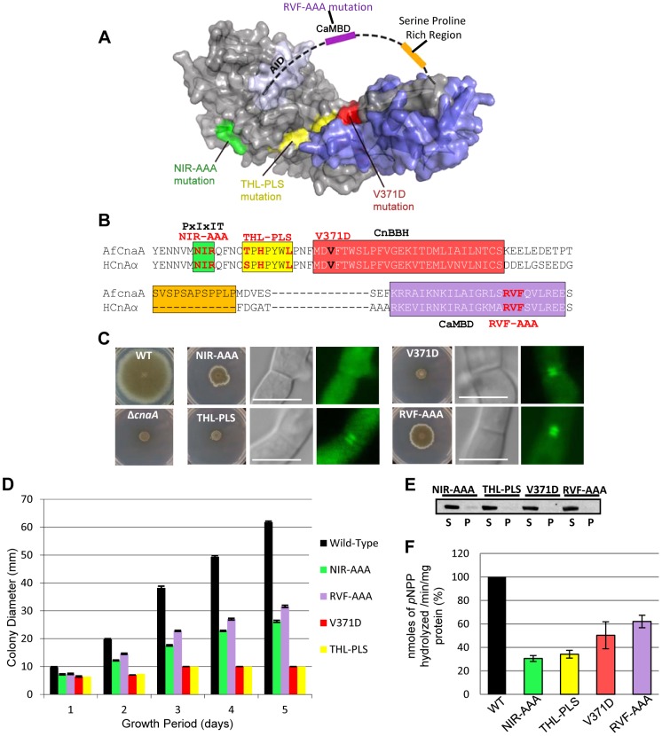
Figure 10. Septal localization of CnaA is independent of Ca2+/calmodulin binding but requires the PxIxIT binding motif.
(A) Homology models for the A. fumigatus CnaA (gray) and CnaB (blue) subunits reveal relative arrangement of subdomains and mutated regions. The Serine Proline Rich Region (SPRR, orange) and calmodulin binding domain (CaMBD, purple) are indicated schematically, as they are likely disordered. (B) Alignment of A. fumigatus CnaA and human CnA-α subunit including the PxIxIT, CnBBH and the CaMBD. (C) Radial growth after 5 days at 37°C on GMM agar. CnaA septal localization after 24 h growth (D) Radial growth is depicted as mean colony diameter in triplicate over 5 days growth. (E) Western detection of CnaA-EGFP fusion proteins using anti-GFP polyclonal antibody and peroxidase labeled anti-rabbit IgG secondary antibody. S and P indicate supernatant and pellet fractions, respectively. (F) Calcineurin activity was determined using p-nitrophenyl phosphate as a substrate. Data from two separate experiments, each with 6 technical replicates are presented as mean ± SD of nanomoles of pNPP released/min/mg protein.

