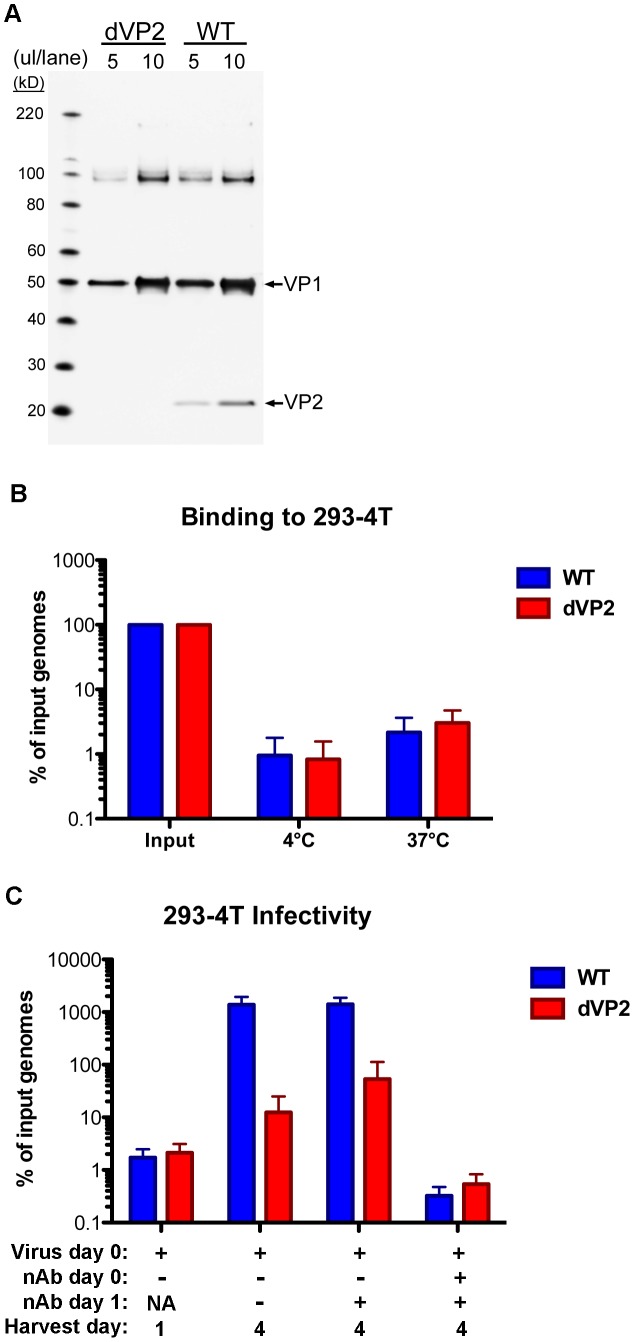
Figure 6. VP2-knockout in native MCV virions.
(A) Purified WT or VP2-knockout mutant (dVP2) MCV virions were analyzed by SDS-PAGE and western blot with a mixture of anti-VP1 and anti-VP2 rabbit sera. (B) Equal amounts of WT and dVP2 virions with equivalent MCV genomic DNA content, were inoculated onto 293-4T cells. Samples were placed at 4°C or 37°C for one hour, and then washed prior to analysis of cell-associated viral DNA as below. (C) Equal amounts of WT and dVP2 virions were inoculated onto 293-4T cells with either neutralizing polyclonal anti-MCV serum (nAb +) or rabbit pre-bleed (nAb −). One sample was harvested the next day (Harvest day 1) while other samples were re-plated +/− neutralizing serum, then harvested day 4 post-infection. The percentage of viral DNA in samples, relative to the amount added in the form of virions, was determined by qPCR. The average of five experiments is shown and error bars represent the standard deviation.

