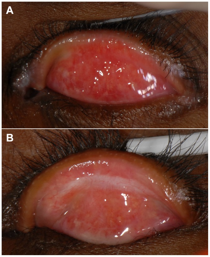
Figure 1. Conjunctivalisation of the lid margin.

1a: The lid margin at baseline showing marked conjunctivalisation; the Meibomian gland orifices are completely surrounded by conjunctival-type surface. 1b: The lid margin of the same participant at the two-year follow-up; the conjunctival surface appears to have receded and the Meibomian gland orifices are surrounded by skin with a normal appearance.
