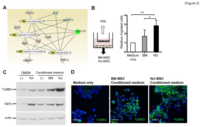
Figure 2. Higher neural induction ability of WJ-MSCs.
(A) An interaction network of secreted factors. Factors that pertained to angiogenesis, vasculogenesis, neurite outgrowth, and neuron migration are connected. Genes in red are abundant in WJ-MSCs and genes in green are abundant in BM-MSCs. (B) Differential chemotaxis effects of WJ-MSCs and BM-MSCs. MSCs cultured in MesenCult® medium were seeded in the lower part of Tranwell plates, while N2a cells were placed in the upper chambers (illustrated in left panel). Migrated N2a cells to the other side of the membrane were stained with Hoechst 33342 and counted. Data are mean ± SD (right panel; *p<0.05, **p<0.01). (C–D) Induction of N2a neural differentiation by MSCs. N2a cells were cultured with MSC conditioned medium, medium only (negative control) or retinoic acid (RA; positive control) for 4 days before the cellular lysates were subjected to Western blotting analysis (C) or the cells were fixed for immunofluorescence staining (D). Neural markers TUBB3 and NEFL were analyzed. Cell nuclei were stained with DAPI. Scale bars: 50 μm.

